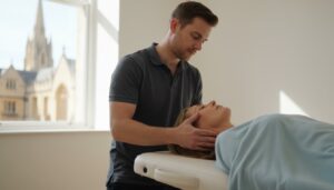
Persistent shoulder pain can be debilitating, affecting everything from simple daily tasks to your overall quality of life. The uncertainty of not knowing the cause, coupled with concerns about potential surgery and long NHS waiting times for scans, can add significant stress. For those seeking a swift and precise answer, a diagnostic ultrasound for shoulder pain offers a modern, effective solution to pinpoint the source of your discomfort without delay, allowing for a clear and immediate path forward.
This guide is designed to provide you with a clear understanding of the entire process. We will explain how this advanced imaging technology works in real-time to assess your tendons, ligaments, and muscles, leading to an accurate diagnosis. By reading on, you will gain the information you need to understand your condition, explore effective treatment options-often avoiding the need for surgery-and take the first confident step towards restoring mobility and achieving long-term pain relief.
Key Takeaways
- Learn how ultrasound provides a clear, real-time view of your shoulder’s soft tissues, accurately pinpointing the source of your discomfort.
- Understand the simple, step-by-step shoulder ultrasound procedure, so you can feel calm and prepared for your appointment.
- Discover how a diagnostic ultrasound for shoulder pain is the key to developing a precise and effective treatment plan tailored to your specific injury.
- See how the detailed results from your scan guide you towards the right therapy to reduce pain and restore long-term mobility.
What is a Diagnostic Ultrasound and Why is it Used for Shoulders?
A diagnostic ultrasound, also known as medical sonography, is a safe and highly effective imaging technique that uses high-frequency sound waves to produce live images of the inside of your body. It is completely non-invasive, involves no radiation, and provides our clinicians with a clear, real-time view of your shoulder’s soft tissues. For those seeking a detailed technical overview of what is medical ultrasound, it is a well-established technology used across many fields of medicine. Its primary role in musculoskeletal care is to examine structures like tendons, muscles, ligaments, and bursae that are often the source of shoulder pain and restricted movement.
To better understand the procedure, this video provides a helpful overview of a shoulder examination:
How Ultrasound Imaging Works: A Simple Explanation
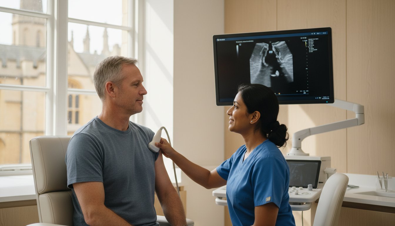
The process is straightforward and painless. It functions much like a ship’s sonar, sending out sound waves that bounce off internal structures. A small, handheld device called a transducer is placed on your skin over a clear, water-based gel. This probe emits and collects the returning sound waves, which a computer then instantly translates into a detailed image on a monitor. The entire process is safe, comfortable, and allows for immediate interpretation by your specialist.
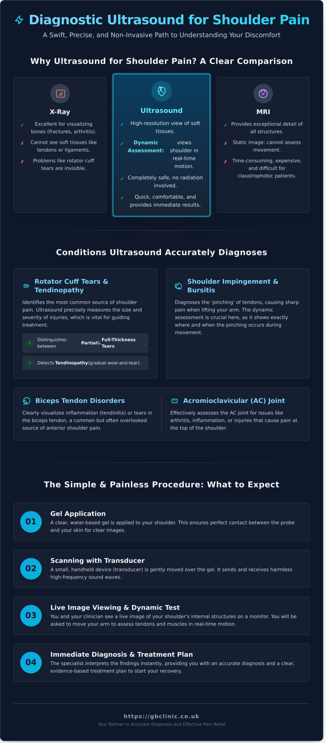
Key Advantages Over X-ray and MRI for Shoulder Issues
While other imaging methods have their place, a diagnostic ultrasound for shoulder pain offers distinct advantages for assessing soft tissue injuries. The key differences are:
- Compared to X-ray: An X-ray is excellent for visualising bones and can identify fractures or significant arthritis. However, it cannot see the soft tissues, meaning common problems like rotator cuff tears, tendinitis, or bursitis are invisible.
- Compared to MRI: An MRI provides exceptional detail of all structures but is a static image, taken while you lie perfectly still. It can also be time-consuming, expensive, and difficult for patients with claustrophobia.
- Ultrasound’s Superiority: Ultrasound provides excellent resolution of soft tissues and, crucially, allows for assessment while the shoulder is moving.
The Power of Dynamic Assessment
The most significant benefit of shoulder ultrasound is its ability to perform a dynamic assessment. This means your clinician can watch the tendons and muscles inside your shoulder as you move your arm. This real-time feedback is invaluable for diagnosing conditions like shoulder impingement, where structures may only become compressed or pinched during specific movements. This unique capability allows for a more precise and functional diagnosis, which is essential for developing the most effective treatment plan to restore movement and reduce pain.
What Shoulder Conditions Can an Ultrasound Accurately Diagnose?
A diagnostic ultrasound for shoulder provides a detailed, real-time view of the muscles, tendons, and ligaments that are often the source of pain and restricted movement. Unlike an X-ray, which primarily shows bones, ultrasound excels at visualising the soft tissues where most shoulder problems originate. This advanced imaging allows our clinicians to make a precise diagnosis and form an effective, evidence-based treatment plan. The scan is particularly adept at identifying:
- Rotator cuff injuries (tears and tendinopathy)
- Shoulder impingement and bursitis
- Biceps tendon disorders
- Acromioclavicular (AC) joint problems
Rotator Cuff Tears and Tendinopathy
The rotator cuff is the most common source of shoulder pain. Ultrasound is highly accurate at detecting injuries to these critical tendons. We can clearly distinguish between a partial-thickness tear (the tendon is damaged but not completely severed) and a full-thickness tear (the tendon is torn all the way through). The scan also identifies tendinopathy, which is the gradual wear-and-tear or degeneration of the tendon tissue, often felt as a deep ache. Crucially, ultrasound allows us to measure the exact size and severity of the injury, which is vital for guiding treatment decisions.
Shoulder Impingement and Bursitis
Shoulder impingement occurs when tendons are ‘pinched’ in the narrow space at the top of your shoulder, often causing sharp pain when you lift your arm. This is frequently associated with bursitis, the inflammation of the bursa (a small, fluid-filled sac that provides lubrication). For more general information on this type of imaging, the Radiological Society of North America offers comprehensive Musculoskeletal Ultrasound patient information. A key advantage of ultrasound is its dynamic capability; we can observe your shoulder in motion to see the exact moment the pinching occurs, confirming the diagnosis with certainty.
Biceps Tendon and AC Joint Issues
The comprehensive nature of a single scan allows us to assess other important structures. The long head of the biceps tendon, which runs through the front of the shoulder, can be a source of pain due to tendinitis (inflammation) or instability (dislocation). Furthermore, the ultrasound can evaluate the acromioclavicular (AC) joint at the top of the shoulder for signs of arthritis, inflammation, or injury. This ability to assess multiple potential pain sources in one efficient appointment makes a diagnostic ultrasound for shoulder an invaluable tool for fast, accurate answers.
The Shoulder Ultrasound Procedure: What to Expect Step-by-Step
Understanding what happens during a medical procedure is key to feeling calm and confident. A diagnostic ultrasound for shoulder pain is a simple, non-invasive process designed entirely around your comfort and achieving a clear diagnosis. The entire appointment, including the scan and discussion of the findings, typically takes around 30 minutes. Below is a step-by-step guide to your examination at our clinic.
Preparing for Your Scan at Our Clinic
One of the key benefits of ultrasound is its simplicity. There is no need for any special preparation, such as fasting or altering your medication schedule. To ensure the process is as smooth as possible, we recommend the following:
- Wear comfortable, loose-fitting clothing that allows easy access to your shoulder area. A vest or loose t-shirt is ideal.
- Bring any relevant documents with you, such as a referral letter from your GP or physiotherapist, and any reports from previous imaging like X-rays or MRI scans.
During the Examination
You will be comfortably seated while the clinician explains each step. First, a small amount of warm, water-based gel is applied to your shoulder. This gel helps the ultrasound probe, or transducer, make secure contact with your skin and allows the sound waves to pass through effectively. The clinician will then gently move the probe over your shoulder, viewing the live images on a monitor.
To get a complete view of the joint structures, you will be asked to move your arm into several different positions. This dynamic assessment is a unique advantage of ultrasound, allowing us to see how your tendons and muscles function in real-time. This methodical approach, as detailed in many a clinical review of shoulder ultrasound, ensures a comprehensive evaluation. The scan itself is not painful, though moving your shoulder into certain positions may cause some familiar tenderness.
Receiving and Understanding Your Results
A significant benefit of a diagnostic ultrasound for shoulder conditions is the immediacy of the results. As the scan is performed, our experienced clinician will explain what they are seeing on the screen. This means you can see the source of your pain for yourself and avoid the anxious wait for a separate report to be written and sent to your doctor.
This instant feedback allows for a clear diagnosis on the same day, enabling you and your practitioner to discuss the next steps and formulate an effective treatment plan without delay. To find out more about how we use this technology for precise diagnoses, learn more about our advanced diagnostic ultrasound scans.
From Diagnosis to Treatment: What Happens After Your Scan?
Receiving a clear diagnosis is the critical first step, but it’s what happens next that truly matters for your recovery. The detailed information gathered from your diagnostic ultrasound for shoulder pain is not just a report; it is the foundation of your personalised recovery journey. At GB Clinic, our integrated approach means you can move seamlessly from diagnosis to treatment, saving you valuable time and ensuring continuity of care under one roof.
Developing a Personalised Treatment Plan
The precise findings from your scan directly inform the most effective course of action. This allows our clinical specialists to create a treatment plan that is targeted specifically to the root cause of your shoulder pain, whether it’s a rotator cuff tear, bursitis, or tendonitis. This evidence-based approach ensures you avoid generic therapies that may prove ineffective, focusing instead on a strategy designed for your unique injury and recovery goals.
Non-Invasive Treatment Options
For many shoulder conditions identified via ultrasound, recovery can be achieved through conservative, non-invasive care. Our expert clinicians can develop a targeted programme of specialised physiotherapy designed to restore function and eliminate pain. Depending on your diagnosis, your plan may include:
- Manual Therapy: Hands-on techniques to improve joint mobility and reduce stiffness.
- Exercise Rehabilitation: A structured programme of exercises to strengthen the shoulder and prevent re-injury.
- Shockwave Therapy: A modern, non-invasive treatment to stimulate healing in chronic tendon issues.
Ultrasound-Guided Injections for Precision Relief
In cases where inflammation or joint degeneration is a key factor, the same ultrasound technology used for diagnosis can be used to guide treatments with pinpoint accuracy. This ensures that medication is delivered directly to the source of the problem for maximum effect. Our clinicians are highly experienced in performing these procedures, which include:
- Guided Steroid Injection: To deliver powerful anti-inflammatory medication directly into an inflamed bursa or joint.
- Hyaluronic Acid (HA) Injections: To lubricate the shoulder joint, reduce pain, and improve mobility in cases of osteoarthritis.
This combination of an accurate diagnostic ultrasound for shoulder assessment followed by a precisely tailored treatment plan gives you the clearest and most efficient path back to a pain-free life. Contact us today to learn how our integrated services can help you.
Why Choose GB Clinic for Your Shoulder Ultrasound in Oxford?
When you are dealing with shoulder pain, receiving a fast, accurate diagnosis is the first and most critical step towards recovery. At GB Clinic, we specialise in musculoskeletal (MSK) conditions and have designed our service to provide clarity and a direct path to effective treatment. Our approach is built on three core principles: specialist expertise, advanced technology, and a fully integrated patient journey.
Expert Clinicians and Advanced Equipment
Your scan will be performed by a highly trained MSK specialist who focuses exclusively on joints, muscles, and soft tissues. This level of specialisation, combined with our modern, high-resolution ultrasound equipment, ensures we capture exceptionally clear and detailed images of your shoulder. The combination of an expert eye and superior technology means your diagnostic ultrasound for shoulder pain provides a precise and reliable foundation for your treatment plan.
A ‘One-Stop’ Approach for Faster Recovery
We understand that your time is valuable and that waiting for separate appointments can be frustrating and delay your recovery. Our Oxford clinic operates as a ‘one-stop’ service, streamlining your care from start to finish. This means you can have your diagnostic scan, receive a clear diagnosis, and begin your recommended treatment all within one dedicated clinic.
- Saves Time: No need to travel between a scanning facility and a separate treatment provider.
- Eliminates Delays: Your treatment plan can be created immediately following your diagnosis.
- Seamless Experience: A smooth and efficient journey from assessment to becoming pain-free.
Integrated Care for Better Outcomes
A diagnosis is only as good as the treatment that follows. At GB Clinic, your diagnostic clinician works in close collaboration with our team of expert physiotherapists. This integrated approach ensures that the insights gained from your scan are directly translated into a highly specific and effective rehabilitation programme. Your treatment is not just based on a report; it’s built on a shared understanding of your unique condition, leading to better, faster results.
Choosing GB Clinic means choosing a dedicated team of MSK specialists committed to your long-term recovery. We provide the clarity you need and the expert, integrated care required to restore shoulder function and help you return to the activities you love. To schedule your appointment or learn more about our services, please contact our Oxford clinic today.
Take the First Step Towards a Pain-Free Shoulder
Shoulder pain should not limit your life, and an accurate diagnosis is the critical first step towards effective recovery. As we’ve explored, a diagnostic ultrasound for shoulder issues provides a clear, real-time view of your tendons, muscles, and ligaments. This clarity removes guesswork, enabling the precise identification of your condition and paving the way for a treatment plan tailored specifically to you.
At GB Clinic, our specialist MSK clinicians use advanced, high-resolution ultrasound technology to ensure diagnostic accuracy. We provide a comprehensive one-stop service where your scan, diagnosis, and personalised treatment plan are managed efficiently under one roof. This approach is designed to give you clear answers and a direct path to relief without unnecessary delays.
Stop guessing about the source of your pain. Take control of your recovery and get the answers you need to move forward with confidence. Book your diagnostic ultrasound scan in Oxford today. We are here to help you restore function and return to a life with improved comfort and mobility.
Frequently Asked Questions About Shoulder Ultrasounds
Is a diagnostic shoulder ultrasound painful?
A shoulder ultrasound is a non-invasive and generally painless procedure. A clear, water-based gel will be applied to your skin, which may feel cool. The clinician will then press a small handheld probe against the shoulder area to capture the images. If your shoulder is already very tender, you may feel mild discomfort from the pressure of the probe, but our specialists are trained to be as gentle as possible to ensure your comfort throughout the scan.
How is an ultrasound different from an MRI for my shoulder?
While both are excellent imaging tools, they use different technologies for different purposes. An MRI uses magnetic fields and provides highly detailed static images of bone, cartilage, and soft tissues. A diagnostic ultrasound for shoulder uses high-frequency sound waves to create real-time images. This allows our clinicians to assess structures like tendons and ligaments while you move your shoulder, providing a dynamic view of the joint’s function that an MRI cannot capture. It is particularly effective for diagnosing rotator cuff tears and tendonitis.
How long will the ultrasound appointment take?
Your complete appointment is designed to be efficient and thorough. The scanning portion of the procedure typically takes between 15 and 20 minutes to complete. We allocate around 30 minutes for the entire appointment to allow for a brief consultation with the clinician before the scan and to discuss the initial findings with you immediately afterwards. This ensures you have a clear understanding of the results without any unnecessary waiting or delays.
Do I need a referral from my GP to have a scan?
No, you do not need a referral from your General Practitioner to book a diagnostic ultrasound scan at our clinic. We offer a direct, self-referral service to provide you with rapid access to diagnosis and treatment. This patient-focused approach helps to avoid long waiting times, allowing you to get the clarity you need to begin your recovery journey as quickly as possible. If you wish, we can share the results with your GP.
Is an ultrasound scan safe?
Yes, ultrasound scans are extremely safe. The technology uses sound waves, not ionising radiation like X-rays or CT scans, so there is no radiation exposure. It is a widely used diagnostic procedure that has no known harmful side effects and is considered safe for all individuals, including pregnant women and patients with medical implants like pacemakers. It is one of the safest and most effective methods for assessing musculoskeletal conditions.
What should I wear for my shoulder ultrasound appointment?
We recommend you wear loose-fitting and comfortable clothing for your appointment. To allow the clinician clear access to the entire shoulder area, it is best to wear a top that can be easily removed or adjusted, such as a vest or t-shirt. You will be provided with privacy to change if necessary. The key is ensuring the sonographer can move the ultrasound probe freely around your shoulder joint and upper arm without obstruction from restrictive clothing.